
Scientists Created a False Fear Memory in a Mouse: What the 2013 Optogenetics Experiment Really Proved
How did MIT scientists create a false fear memory in a mouse? Explore the 2013 optogenetics experiment, controls, DG versus CA1 results, and human limits.
Contents
- The experiment in one scene: a room that became frightening without a shock
- What counts as a false memory when the subject cannot describe it?
- Why the hippocampus—and why the dentate gyrus in particular?
- How optogenetics made the experiment possible
- The three environments: a step-by-step reconstruction
- Freezing is a meaningful measurement, but not mind reading
- A crucial comparison most simplified articles leave out: DG versus CA1
- How did scientists rule out ordinary generalized fear?
- Did the false association erase the genuine memory of context B?
- False memories in mice and humans: similar theme, different evidence
- Could the discovery lead to treatments for trauma or PTSD?
- The next decade: memory engrams became more complicated, not less
- Six viral claims compared with the evidence
- Why the study changed the memory debate
- Conclusion: stranger—and more limited—than science fiction
- Primary sources and further scientific reading
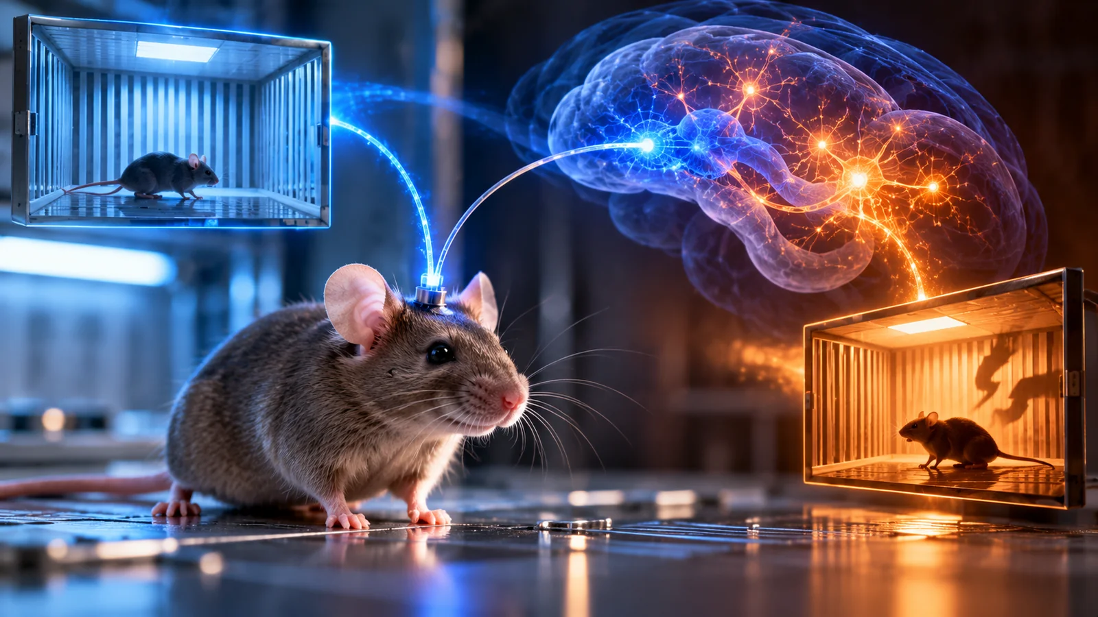
A mouse entered a room where nothing bad had ever happened to it. Yet, when researchers returned the animal to that same room days later, it froze as though danger had occurred there. The twist was not that the mouse had forgotten the location of a shock. It had never received a shock in that room. Scientists at the Massachusetts Institute of Technology had artificially reactivated a neural representation of the safe room while the mouse experienced a real unpleasant event somewhere else. The result, published in Science in July 2013, became one of the most striking demonstrations of how a memory-related association can be altered at the level of identified brain cells.
The experiment is often described as “implanting a false memory.” That phrase captures the surprise, but it can also distort the science. The researchers did not download a detailed scene into a brain, record a mouse’s thoughts, or demonstrate that they could implant a human childhood memory. They showed that, under a carefully controlled animal protocol, they could establish an incorrect association between a previously neutral context and a fear response. This distinction is the key to the study’s value—and to understanding what its findings do not license us to claim. This article examines the original Ramirez and colleagues paper, related research, and subsequent reviews rather than reporting any experiment performed by Horus Valley.
The experiment in one scene: a room that became frightening without a shock
Imagine two distinctly different experimental chambers. In chamber A, a mouse explores freely: it senses the walls, floor, lighting, odors, and geometry. No foot shock occurs there. In chamber B, encountered later, the mouse receives mild electrical foot shocks as part of a laboratory fear-conditioning protocol. Normally, it would be reasonable for the animal to associate B with an unpleasant experience. But the researchers added an intervention during those shocks. They used precisely timed light stimulation to activate a set of hippocampal cells that had been active when the mouse explored A the day before.
After that manipulation, the mouse was returned to A. Instead of behaving as if the setting were neutral, it exhibited increased freezing, a standard behavioral measure of conditioned fear in rodents. This was the central result: the actual shock had taken place in B, but a fear-related response emerged in A. The researchers had made the remembered representation of one context participate in an association formed during a different experience. MIT’s contemporaneous account explains the day-by-day design; the published paper provides the evidence and experimental comparisons.
It is tempting to describe this as rewriting the animal’s past, but the statement needs precision. The historical fact of the shock’s location did not change, and the scientists did not establish that the mouse mentally pictured a detailed fictional episode. They linked an already acquired neural representation of a place to a real aversive event experienced elsewhere. That is an experimentally induced false contextual fear association, not proof of arbitrary memory editing. The difference matters because an accurate explanation should be at least as interesting as a sensational one.
What counts as a false memory when the subject cannot describe it?
Human false-memory research often depends on what a participant reports remembering or believing: a suggested childhood incident, a misleading detail inserted after an event, or a source of information mistaken for personal experience. A mouse cannot supply such a verbal account. The neuroscientists therefore turned the question into a behavioral experiment: if a representation of a safe place is activated while the animal learns fear elsewhere, will that safe place later provoke fear in a context-specific manner? They could observe and compare that outcome even though they could not ask the mouse what it thought had happened.
This introduces three different levels that popular accounts frequently collapse. First is the external event—where the actual shock occurred. Second is the neural representation—the patterns and cellular ensembles engaged when a particular context is experienced or recalled. Third is the measured behavior—freezing or another fear-related response. The 2013 study connected these levels causally under controlled conditions. But a mismatch between event and behavior is not the same as direct access to subjective experience. No instrument in the study read out a first-person narrative in the mouse’s mind.
Memory is also not a static video file that can simply be opened and replayed. Recollection involves reconstruction and updating; experiences are represented across interacting circuits, and their significance can change with subsequent learning. A 2025 cross-species review of memory modification describes adaptive and maladaptive updates in humans and rodents while emphasizing important differences between methods and species. The mouse experiment becomes easier to understand when placed within this broader view: associations between components of experience can be altered, but doing so does not imply limitless control over all memories.
Why the hippocampus—and why the dentate gyrus in particular?
The hippocampus is a brain region deeply involved in forming relationships among the places, events, and temporal elements that make up experience. It is not a single vault storing every memory, nor does it function independently of the cortex, amygdala, and other systems. For a question involving fear attached to a particular place, however, hippocampal circuits are especially useful. Within the hippocampus, the dentate gyrus (DG) helps distinguish among contextual representations that share features. The researchers targeted cells whose activity became associated with exploration of one environment and asked whether those cells could later be used to influence learning.
The relevant concept is a memory engram. In modern neuroscience, an engram refers broadly to enduring changes in a population of cells and their connections that help encode, retain, or retrieve aspects of an experience. An engram is not the same as one neatly labelled neuron holding a complete scene. Different cells and regions contribute to different parts of a memory, and their activity can change across time. A 2024 review of engram research underscores the distributed nature of these representations and the limits of methods that monitor or manipulate one cell class or region at a time.
In earlier work, the MIT group had identified neurons recruited during a real fear-conditioning experience and shown that later stimulation could evoke fear-related freezing in a different setting. That was an important step from correlation toward causation: manipulating candidate engram cells could affect memory-related behavior. The new 2013 question was more ambitious. Could an engram associated with a neutral, unshocked context become linked to fear when reactivated during a shock delivered elsewhere? The answer, in particular experimental conditions, was yes. That is why this study was about creating a new association rather than simply replaying an old one.
How optogenetics made the experiment possible
The technique is called optogenetics, a combination of genetic targeting and optical control. In the study, selected neurons could express channelrhodopsin-2 (ChR2), a light-sensitive protein that can drive neuronal activity when exposed to appropriate light. A system linked to activity of the immediate-early gene c-fos, together with controlled periods of doxycycline exposure, allowed researchers to label cells recruited while an animal explored a specific environment. Implanted optical fibers could then activate the labelled population during a later experimental phase.
There are two distinct processes here. Labelling identifies neurons active during the chosen contextual experience, making a defined group available for later intervention. Reactivation then stimulates that group when an unrelated experience takes place. This is not what happens when an ordinary light shines through someone’s scalp. For background on the method rather than this particular experiment, see Horus Valley’s guide to optogenetics and the 2026 Nobel Prize. The procedure required a specialised genetically engineered animal system, viral genetic constructs, optical hardware, and validation that the stimulated neurons actually responded. Nothing in the published protocol resembles a consumer light therapy, a hypnosis technique, or a noninvasive way to program someone’s thoughts.
Nor was the technology a camera capturing the memory itself. The researchers observed and manipulated neural activity correlated with a known experience, then assessed its causal role in behavior. The distinction between tracking a neuronal population and decoding an entire episode matters. Even highly sophisticated activity tagging reveals only selected biological features of a complex and distributed memory system. Later literature discusses how engrams change over time, how inhibitory and excitatory networks interact, and why claims about fixed one-to-one memory maps remain oversimplified.
The three environments: a step-by-step reconstruction
| Stage | What happened | What the researchers needed to establish |
|---|---|---|
| Preparation | Targeted neurons were equipped to express a light-sensitive protein during a defined activity window | Cells engaged by a particular experience could later be reactivated |
| Context A | The mouse explored a novel chamber without receiving foot shocks | A neural representation of a safe context could be labelled |
| Context B | On a later day, the animal received foot shocks while labelled A cells were optically activated | The internal representation of A could coincide with real fear learning in B |
| Return to A | Researchers measured freezing in the original, unshocked chamber | Fear associated with A would indicate a false contextual association |
| Control context C | The mouse was tested in another distinct environment | The response should be specific rather than a generalized fear of all places |
Context A is the setting at the heart of the false-memory claim. If shocks had been delivered there, later freezing would be ordinary learning rather than a misleading association. Context B is where the aversive event actually occurred; the procedure did not manufacture a fear response without any aversive input. Context C served a different purpose: checking whether the response was tied to the reactivated representation of A rather than to undifferentiated distress or fear of any unfamiliar location.
A useful way to visualize the causal hypothesis is: activity representing A + shock occurring in B → later fear response to A. That is a conceptual summary, not an equation that predicts a fixed success rate. The biological effect depended on how neurons were tagged, the specific area targeted, stimulus timing, experimental controls, and the animals’ behavior. A detailed researchers’ account published in 2014 walks through those procedures and compares the behavioral and neural evidence.
Freezing is a meaningful measurement, but not mind reading
In rodent fear-conditioning research, freezing describes a relative absence of movement that can occur when an animal anticipates danger. Under controlled conditions, the amount of freezing serves as an established behavioral indicator of learned fear. However, a reader should resist converting that measure directly into a statement such as “the mouse vividly remembered a fictional electric shock.” The study was not equipped to recover subjective pictures, language, or autobiographical descriptions from the animal. It measured behaviors and neural responses that support a narrower inference.
That is why control experiments mattered. The researchers did not merely notice one mouse standing still. They compared contexts, stimulation conditions, and cellular targets. They also investigated whether light-based activation could trigger a fear response in another setting and whether relevant downstream circuits became engaged. Such results support the idea that a functional fear association had been formed, rather than a nonspecific motor disturbance produced by the experimental apparatus. Still, similarity in measured neural pathways does not establish that genuine and false memories are identical in every respect.
Some articles attempt to convert the study into a global success percentage or suggest that all experimental mice responded in exactly the same way. That would be inappropriate without specifying the particular figure, outcome definition, protocol, and control condition. A freezing percentage measures time spent in a behavior during an observation window; it is not a percentage of memory content successfully written into a brain. The original research should be read in terms of group comparisons, conditions, and statistical interpretation, not as a market-style effectiveness claim.
A crucial comparison most simplified articles leave out: DG versus CA1
The hippocampus is not a uniform block of memory tissue. The scientists tested manipulations involving CA1, another hippocampal subregion, alongside those involving the dentate gyrus. Under the same behavioral and stimulation parameters described for this experiment, activating tagged CA1 cells did not produce the corresponding increase in freezing in context A observed with DG tagging. This is an important constraint. It argues against the idea that stimulating just any memory-related neurons during fear conditioning automatically creates the same false association.
The correct conclusion is not that CA1 has no role in memory. Other tasks, protocols, and experiments implicate CA1 in different aspects of memory processing. What this particular comparison shows is that the circuit, the targeting method, and the precise experimental conditions matter. The authors did not discover a universal “false memory switch.” Instead, they demonstrated a specific causal manipulation within a carefully defined network. That is more informative than saying simply that scientists “turned memories on with lasers.”
| Comparison | Dentate gyrus (DG) | CA1 under these study conditions | What the comparison establishes |
|---|---|---|---|
| Context-related activity tagging | Performed | Performed | Cells from both regions could be selected |
| Optical reactivation during fear learning | Performed | Performed | Intervention was not limited to one region |
| Extra freezing in unshocked context A | Seen in the experimental condition | Corresponding increase was not observed | The outcome depended on target and protocol |
| Broad claim that would be unjustified | DG contains every detail of every memory | CA1 is irrelevant to memory | Neither follows from this experiment |
How did scientists rule out ordinary generalized fear?
Suppose the mouse merely became frightened by the laboratory procedure and therefore froze in any later room. Then returning to A would tell us relatively little about a specific false association. The study addressed that possibility by testing a novel control context C and comparing appropriate groups. The experimental animals showed an enhanced response to A, not the same pattern across every environment. The context specificity strengthened the claim that the tagged representation of A was selectively linked with fear learning during B.
Other comparisons asked whether illumination itself, cellular activation unrelated to the intended memory, or differences in the target region could explain the outcome. Controls involving animals without the functional light-sensitive protein and manipulation of a different hippocampal region helped address those alternatives. The 2014 detailed scientific review also describes an active fear response elicited when the tagged DG cells were reactivated in another context; downstream activity related to natural fear recall provided an additional line of evidence.
No individual control rules out all possible uncertainties, but together they form a coherent causal argument. This is the scientific heart of the paper and a major opportunity for responsible popular explanation. The extraordinary aspect was not the spectacle of a mouse stopping movement. It was the pattern of outcomes across conditions deliberately designed to distinguish a context-specific learned association from nonspecific fear or optical side effects.
Did the false association erase the genuine memory of context B?
Not necessarily, and “erasure” would be a misleading description. The animals could also display fear linked to the actual shock location, B. The researchers discussed potential competition between the false and genuine fear memories: when an internally reactivated contextual cue becomes paired with the same aversive event as the present physical environment, the two associations may affect each other’s expression. This fits into a broader literature on how cues compete and cooperate during learning. It does not mean scientists selected one memory file and permanently replaced another.
Consider a deliberately imperfect everyday analogy. If someone experiences a frightening moment while a familiar song is playing, that song might later evoke unpleasant feelings even in a safe setting. In the mouse study, the analogue of the familiar song was not played externally; a neural ensemble tied to the harmless context was activated directly. The analogy helps explain association, not the technical method, and it should never be presented as an independent experiment proving that ordinary thoughts or sounds can reproduce the MIT result.
We should also distinguish changing the emotional significance of a cue from manufacturing a complete history. A room can become threatening in a learned response without every feature of the animal’s representation of the room changing. Equally, demonstrating one neural mechanism for contextual fear does not establish that all traumatic memories, everyday autobiographical events, and complex beliefs can be edited with the same intervention. The scale and kind of information being measured matter.
False memories in mice and humans: similar theme, different evidence
Research on human memory distortion includes misinformation effects, source confusion, suggestion in interviews, and the formation of false autobiographical beliefs or recollections under certain conditions. Such studies typically rely on participants’ reports, recognition tests, confidence judgments, and source-monitoring tasks. The 2013 mouse study used a very different intervention and outcome: genetic tagging, optogenetic stimulation, fear conditioning, and behavioral measurement. Both concern the reconstructive nature of memory, but they answer distinct questions and cannot be treated as interchangeable demonstrations.
A notable human literature concerns the classic “lost in the mall” paradigm, in which fabricated childhood-event information is supplied alongside real details. A preregistered replication reported in 2023 found evidence of false-memory reports in some participants; subsequent analysis published in 2024 questioned how well some classifications corresponded to participants actually remembering the event’s central features. Presenting both studies conveys a more informative lesson than quoting one memorable percentage as a universal human susceptibility rate.
The results of neither literature justify saying that lasers can make someone confess to a fictional childhood event. Human remembering is intertwined with language, beliefs, social information, and self-narrative; rodent contextual conditioning offers a controlled model of one important component. A careful comparison highlights a shared caution—confidence and emotional force do not guarantee historical accuracy—without claiming that the mouse intervention demonstrates an immediate clinical or investigative capability in people. For the broader psychology of source confusion and misleading suggestions, see the previously published Horus Valley article How False Memories Form. This article focuses instead on the specific 2013 mouse experiment.
| Dimension | MIT mouse experiment, 2013 | Human false-memory studies |
|---|---|---|
| Target | Context representation and fear association | Recollection, belief, or details of experienced events |
| Intervention | Activity tagging and optogenetic reactivation during conditioning | Suggestion, misinformation, interviews, or other cognitive methods |
| Outcome | Freezing and selected neural measures | Participant reports, memory judgments, and related tests |
| Supported conclusion | False contextual fear association can be induced in mice under experimental conditions | Some memories and beliefs can be distorted or fabricated under studied conditions |
| Unsupported leap | Arbitrary upload of human memories | Demonstration of equivalent optogenetic mechanism in humans |
Could the discovery lead to treatments for trauma or PTSD?
The result has inspired legitimate questions about fear, memory updating, and the circuits that give past experiences their emotional charge. It may help researchers think about ways to understand maladaptive learning and how associations are retrieved or modified. That scientific relevance is not the same as an approved therapy. The paper did not test treatment for human post-traumatic stress disorder, did not establish that a patient’s traumatic memory could be replaced with a pleasant one, and did not justify clinical claims about targeted memory implantation.
Translational challenges are substantial. A complex human traumatic memory is not equivalent to an experimentally trained mouse associating a specific chamber with a foot shock. Human engrams are embedded in distributed networks, and the intervention used in the animal work required invasive optical hardware and genetic manipulation. Clinical adoption would require evidence of safety, specificity, durability, real-world benefit, and ethical acceptability—not only a striking laboratory demonstration. Recent reviews continue to emphasize that many foundational engram findings arise from animal models with limited direct transfer to humans.
Someone distressed by intrusive memories should not interpret this research as a do-it-yourself technique or as a reason to reject established mental-health care. Psychological treatments and other clinical approaches may be helpful for some conditions when selected by qualified professionals. That is a separate evidence base. The responsible public-health message is to understand why scientists study fear memories without suggesting that experimental optogenetic procedures are available, safe, or appropriate for individual patients.
The next decade: memory engrams became more complicated, not less
After 2013, studies of memory engrams expanded beyond the idea of finding cells associated with a single event. Researchers investigated how memories become linked if they occur near one another in time, how representations reorganize across brain regions, how cell populations change, and how the same neural networks can support both retention and updating. A 2024 Nature Reviews Neuroscience article discusses overlapping engrams and the preservation of distinct memory identities, offering a more nuanced framework than the fantasy of one uniquely labelled neuron for every life event.
A 2025 review across human and rodent research emphasizes the continuing process of memory updating and the diverse molecular and circuit mechanisms that may contribute. A 2024 review of the engram concept argues that single methods capture only parts of the overall process: populations include excitatory and inhibitory neurons, large- and small-scale circuits, and representations that can drift over time. These developments strengthen the importance of the early causal experiment while placing tight boundaries around oversimplified interpretations.
Even what a memory is “made of” remains actively debated at several levels. A 2025 survey of neuroscientists found broad support for roles of neuronal connectivity and synaptic strength but no simple consensus on the exact structural details that would completely specify a long-term memory. Such disagreement is not evidence that neuroscience has failed. It reflects how many explanatory levels sit between an identified cell ensemble and the lived experience of remembering one’s life.
Six viral claims compared with the evidence
| Viral claim | Evidence-based assessment | More accurate explanation |
|---|---|---|
| “Researchers uploaded a memory into a mouse.” | Misleading shorthand | They reactivated an existing context-associated ensemble during fear learning |
| “The mouse was shocked in the safe room.” | False | Foot shocks took place in B, while fear was later observed in A |
| “Any laser can implant thoughts.” | Unsupported | Specialised genetic, viral, optical, and behavioral methods were required |
| “It was just generalized fear.” | Incomplete | Context and control comparisons supported a specific learned association |
| “Scientists can now edit human memories at will.” | Unsupported | The demonstrated manipulation was in rodents under experimental conditions |
| “False and real memories are identical.” | Overstatement | Some measured behavioral and downstream neural responses overlapped |
There is a useful five-question reading method for any sensational neuroscience headline: Which species was studied? What intervention was performed? What was actually measured? What control condition was used? What conclusion did the authors support? If the article cannot answer those questions, its promise of a spectacular discovery is likely outpacing the evidence. This is also why a responsible piece should separate a vivid artist’s illustration from actual images obtained in the laboratory.
Why the study changed the memory debate
For decades, neuroscientists could observe areas that became active during learning or recall, but observing co-occurrence is not the same as demonstrating causation. Ramirez, Liu, and colleagues were able to target cells recruited by a neutral context and activate them at a critical moment of new learning. Later, the animal’s response to that neutral context changed. In other words, the manipulation did not just evoke an established memory; it influenced what became associated with that memory-related representation. The experimental timing was part of the mechanism, not a minor procedural detail.
The paper also offers a lesson in how to interpret apparently contradictory evidence. The physical world says a shock occurred in B. The later behavior says A had acquired fear-related significance. No one needed to suppose that the room itself changed or that a fictional event had materialised. The mismatch emerged because an internal representation of one place had been artificially coupled to the emotional significance of another. This is a concrete example of how a brain’s representation of the past can diverge from the past itself.
A story like this can be sensationalized in a few words, but its enduring appeal is scientific rather than cinematic. The authors tested a difficult question with controls that separated competing explanations and linked a definable neural population to a meaningful change in behavior. They also left fundamental questions open: which networks support distinct aspects of false memories, how do associations change with time, what parallels exist across species, and what could ever be safe or useful in human medicine?
Conclusion: stranger—and more limited—than science fiction
The 2013 Science paper did not show that scientists can manufacture anyone’s memories from scratch. It demonstrated a more precise and experimentally powerful result: optogenetic reactivation of hippocampal neurons associated with an unshocked chamber, timed to coincide with fear conditioning in a separate chamber, led mice to show increased fear in the original environment. Behavioral controls and additional neural measures supported the interpretation of a context-specific false fear memory, within the constraints of a mouse model.
The central insight remains important more than a decade later. Memory is neither an untouchable archival recording nor an infinitely editable document. Its components can be reactivated, linked, and updated through biological processes whose details researchers are still uncovering. The most compelling account of the experiment is therefore also the most careful one: a remarkable causal link between a neural representation, a new association, and behavior—not a demonstration of human mind control.
Primary sources and further scientific reading
- Ramirez et al., Science (2013): Creating a False Memory in the Hippocampus — original experimental paper.
- MIT News (2013): Neuroscientists Plant False Memories in the Brain — institutional account of the experiment and its interpretation.
- Liu, Ramirez & Tonegawa, Philosophical Transactions B (2014) — detailed scientific overview of methods, controls, and results.
- Lopez et al., Neuroscience & Biobehavioral Reviews (2024) — review of memory engrams and unresolved questions.
- Brunswick et al. (2025): Memory Updating and Distortion in Humans and Rodents — cross-species review.
- Choucry et al., Nature Reviews Neuroscience (2024) — engram linking and identity.
You may also like
 Psychology
Mental Fatigue vs. Burnout: How to Tell the Difference and What Actually Helps
Mental fatigue or burnout? Compare symptoms, workplace causes and depression red flags with a practical, non-diagnostic guide grounded…
 Psychology
Can Mice Inherit Their Fathers’ Fear? The Odor Experiment Behind a Controversial Epigenetics Claim
A 2014 mouse experiment linked fathers’ odor–shock learning to odor sensitivity and sensory changes in descendants. Here is…
 Psychology
The Marshmallow Test Revisited: Can Waiting for a Treat Really Predict a Child’s Future?
Does the marshmallow test predict success? Compare original research with the 2013 trust experiment, 2018 replication, 2020 reanalysis…