
Dinosaur Blood in a 115-Million-Year-Old Fossil? What the Evidence Shows
A Brazilian dinosaur fossil contains heme signatures and cell-like structures. See how researchers tested the evidence, why it matters, and what remains debated.
Contents
- What did the dinosaur-blood study actually find?
- How did the team test the claim?
- Why a cell-like shape alone would not be enough
- How could traces survive for 115 million years?
- Why are some scientists cautious?
- Which dinosaur supplied the fossil?
- Does this bring dinosaur cloning closer?
- What to watch for next
- Sources and image credits
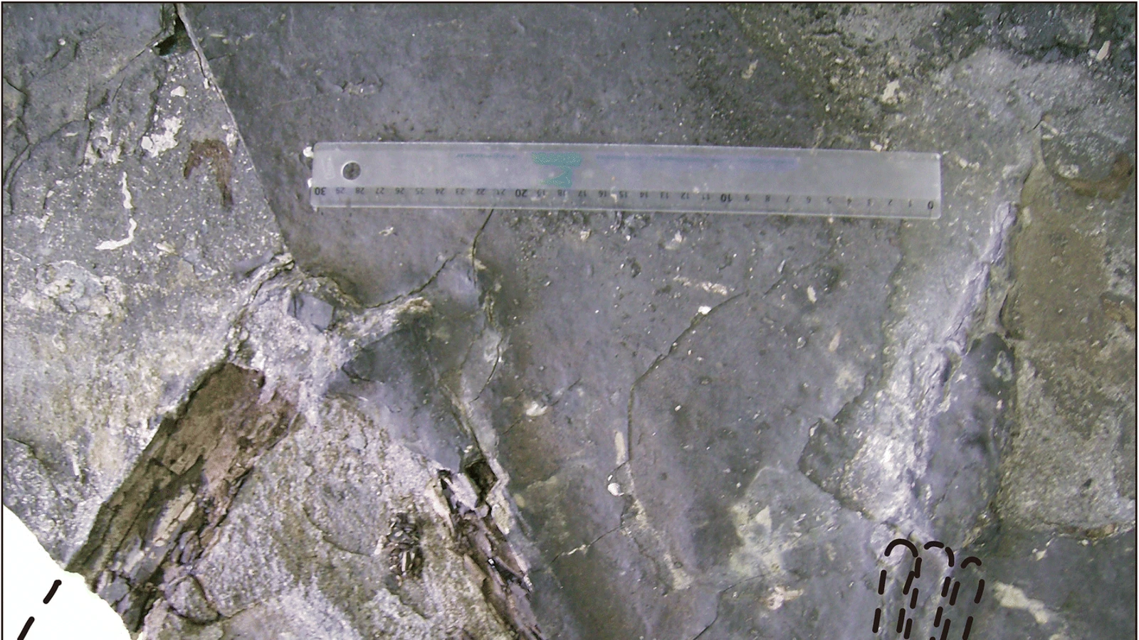
Researchers have reported blood-derived molecules inside mineralized structures interpreted as red blood cells in a 115-million-year-old Brazilian dinosaur bone. The find concerns chemical traces and fossilized microstructures. It does not establish living cells, liquid blood, or recoverable dinosaur DNA.
The study, published in PNAS on 2 November 2026 and announced by Curtin University on 3 November, examined Aratasaurus museunacionali. Its strongest point is the combination of microscopic location and molecular chemistry. The interpretation of the cell-like structures is also attracting scientific scrutiny.
What did the dinosaur-blood study actually find?
The published abstract describes oval microstructures restricted to vascular canals, the channels within bone. Analyses identified heme-related compounds in the fossil and associated heme signals with the structures. Heme is the iron-containing component used by hemoglobin in oxygen transport.
The authors interpret this combination as evidence of blood-derived biomolecules within mineralized red blood cells. Their wording matters: a molecule associated with blood, a cell-shaped structure, and a functioning blood cell are different kinds of evidence.
| Term in the coverage | What it means here |
|---|---|
| Blood-derived molecules | Chemical traces associated with heme |
| Cell-like structures | Microscopic shapes interpreted by the team as red blood cells |
| Mineralized preservation | Structures associated with minerals rather than unchanged living tissue |
| Dinosaur DNA | Not established by these findings |
How did the team test the claim?
The study combined several methods. High-performance liquid chromatography–mass spectrometry identified parent and chemically altered heme B in bone extracts. Raman spectroscopy examined molecular signals at specific locations. Transmission electron microscopy and elemental analyses investigated the microstructures and their mineral associations.
The comparison material is important. The surrounding rock had a different porphyrin signature, associated with chlorophyll and bacteriochlorophyll. That difference supports the authors’ argument that the bone’s signal came from the fossil rather than simply matching the surrounding sediment. It is evidence for their interpretation, not a claim that every possible contamination route has been eliminated.
The participating Brazilian institution, Universidade Regional do Cariri (URCA), separately explains that the team compared the bone and its enclosing sediment. It identifies heme B and a related compound, 3,8-diethyl heme B, and explicitly cautions that the results do not mean intact blood cells survive inside the fossil.
Why a cell-like shape alone would not be enough
In her interview with ScienceAlert, lead researcher Kliti Grice emphasizes the need to combine microscopy with chemical evidence. The team considered alternative origins for the structures, including microbial material and mineral features. The argument therefore depends on several observations agreeing, rather than one striking microscope image.
For readers evaluating a future headline, ask three questions: Where was the signal found? What identified the molecule? What comparison was used? A shape establishes appearance; a chemical test establishes composition; a spatial analysis tests whether the chemistry and shape occur together. Those questions help explain why this study is more specific than a claim that “something red” was seen in a bone.
How could traces survive for 115 million years?
According to Curtin University’s announcement, the structures were associated with hausmannite, a manganese oxide mineral. The proposed preservation pathway involves rapid burial in an oxygen-poor, microbially rich setting, with mineralization helping stabilize delicate biological features and associated compounds.
This is a reconstruction of the preservation process. It does not mean a pristine blood cell simply sat unchanged for the entire interval. Grice’s explanation to ScienceAlert instead describes biological material interacting with its geochemical surroundings, potentially leaving a mineralized record of something normally lost quickly.
A useful distinction is between survival of information and survival of a working cell. A fossil can retain chemical or structural clues after biological function has ended. Understanding which clues remain, and how they were altered, is the research problem.
Why are some scientists cautious?
The independent experts interviewed by ABC Science did not all assess the cell interpretation the same way. Mauricio Barbi considered the mineralized structures consistent with red blood cells but stressed that their identification still involves interpretation. Mary Schweitzer questioned whether the reported shapes and size convincingly establish red blood cells, while accepting the plausibility of heme and collagen-related material in the bone.
These reactions concern the strength of particular interpretations. They should not be flattened into either “everyone agrees” or “the discovery has been disproved.” The research team presents multiple lines of evidence; other specialists are examining whether those lines uniquely identify the structures. Replication and additional specimens can test how broadly the result holds.
Which dinosaur supplied the fossil?
Aratasaurus museunacionali was already known before this molecular study. Its 2020 scientific description documented an incomplete, articulated right hind limb from northeastern Brazil, including parts of the femur, tibia and foot. The specimen was a juvenile, with an estimated body length of approximately 3.12 metres.
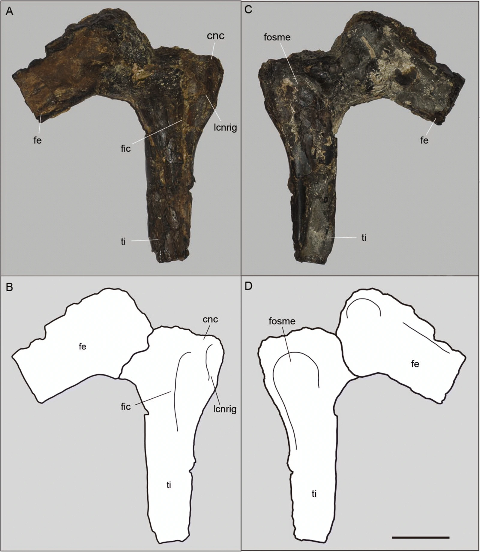
URCA reports that the fossil was found in 2008 in Santana do Cariri, Ceará. The new work adds molecular evidence to a specimen previously investigated anatomically. “New discovery” here describes a new finding within an existing fossil, rather than a new dinosaur dug up in November 2026.
Does this bring dinosaur cloning closer?
This study does not demonstrate recoverable dinosaur DNA. Heme chemistry is not a genome. ABC reports that DNA testing of the samples was negative. The reported structures are mineralized remnants, not viable cells that could be grown in a laboratory.
The realistic opportunity is to investigate fossil preservation and potentially ancient physiology. Curtin’s researchers see possibilities for studying oxygen transport, metabolism and molecular longevity. Those are future research directions, rather than a completed reconstruction of this dinosaur’s metabolism from the present findings.
What to watch for next
- Independent analyses of additional specimens and geological settings.
- Further tests of whether chemical signals consistently align with the proposed cell structures.
- Experiments examining how mineralization preserves or replicates biological features.
In her ScienceAlert interview, Grice identifies replication as an immediate priority. Existing museum collections may contain material worth re-examining with complementary techniques. For another example of how fossil evidence can change an interpretation, see our guide to new research on the timing of mass extinctions.
Sources and image credits
- Grice and colleagues — PNAS research article, published 2 November 2026; DOI: 10.1073/pnas.2614248123
- Curtin University — research announcement, 3 November 2026
- URCA — participating institution’s report, 3 November 2026 (Portuguese)
- Sayão and colleagues — original Aratasaurus description, Scientific Reports, 2020
- ABC Science — interviews with the lead researcher and independent specialists, 3 November 2026
- ScienceAlert — interview with lead researcher Kliti Grice, 3 November 2026
Sources reviewed on 3 November 2026. The PNAS abstract, significance statement and publication record were available; the full main text was access-restricted. The account above uses those public sections, participating institutions’ reports and attributed interviews.
Images: Juliana Manso Sayão, Antônio Álamo Feitosa Saraiva, Arthur Souza Brum, Renan Alfredo Machado Bantim, Rafael Cesar Lima Pedroso de Andrade, Xin Cheng, Flaviana Jorge de Lima, Helder de Paula Silva and Alexander W. A. Kellner, Scientific Reports (2020), Figures 4 and 5, CC BY 4.0. The cover is a 16:9 crop of the pre-preparation fossil photograph; the in-text anatomy figure is resized and converted to WebP. These are 2020 specimen images, not micrographs from the 2026 heme study. Original scientific labels are retained in the anatomy figure.
You may also like
 Science & Discoveries
UK November Cold Snap: What the Met Office Forecast Says
What the Met Office forecast says about the UK November cold snap, possible northern wintry showers, frost and…
 Science & Discoveries
Altermagnetism Explained: What the New Evidence Shows
What is altermagnetism? Understand the new MnTe experiment, how it differs from other magnetic orders, and what the…
 Science & Discoveries
White Christmas 2026: What Can We Actually Predict?
Can we predict a white Christmas in 2026? Learn to read NOAA’s historical snow map, understand the winter…